
การติดตามความดันของลูกโป่งท่อหายใจระหว่างผ่าตัดหลังจากขยายลูกโป่งด้วยการทดสอบการรั่วไหลด้วยความดันบวกในผู้ป่วยเด็กที่เข้ารับการผ่าตัดหัวใจแบบเปิด
Article Sidebar
Main Article Content
บทคัดย่อ
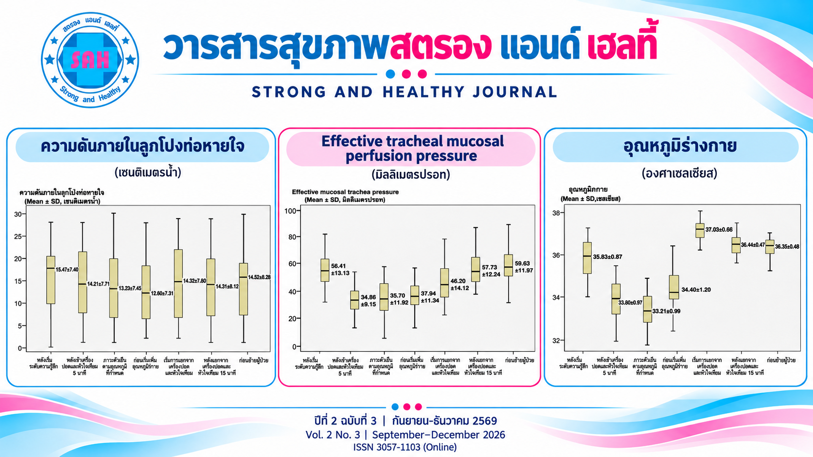
การใช้ท่อหายใจชนิดมีลูกโป่งในผู้ป่วยเด็กที่ผ่าตัดหัวใจช่วยลดการรั่วของลมและภาวะแทรกซ้อนได้ดี แต่การใช้เครื่องปอดและหัวใจเทียม (CPB) ทำให้เกิดการเปลี่ยนแปลงซึ่งอาจส่งผลต่อความดันในลูกโป่งท่อหายใจ ความดันลูกโป่งที่ไม่เหมาะสมอาจทำให้เกิดอันตรายต่อผู้ป่วยได้ มีวัตถุประสงค์เพื่อศึกษาการเปลี่ยนแปลงของความดันลูกโป่งท่อหายใจและปัจจัยที่เกี่ยวข้องระหว่างการผ่าตัดหัวใจโดยใช้เครื่องปอดและหัวใจเทียมในผู้ป่วยเด็ก การศึกษาเชิงสังเกตแบบไปข้างหน้าในผู้ป่วยเด็กอายุต่ำกว่า 8 ปี จำนวน 40 ราย ที่เข้ารับการผ่าตัดหัวใจแบบเปิด หลังใส่ท่อหายใจได้เติมลมในลูกโป่งด้วยวิธีทดสอบการรั่วแบบแรงดันบวก และติดตามวัดความดันลูกโป่ง ในช่วงเวลาต่างๆ ตั้งแต่เริ่มระงับความรู้สึกจนถึงก่อนย้ายผู้ป่วยออกจากห้องผ่าตัด วิเคราะห์ข้อมูลด้วยสถิติ Repeated Measures ANOVA
ผลการศึกษาพบว่าความดันลูกโป่งเริ่มต้นเฉลี่ยอยู่ที่ 15.47 ± 7.40 เซนติเมตรน้ำ พบว่าความดันลูกโป่งลดลงอย่างมีนัยสำคัญทางสถิติ (p < 0.05) ในช่วงการใช้เครื่องปอดและหัวใจเทียม โดยต่ำสุดในช่วงก่อนเริ่มเพิ่มอุณหภูมิกาย (12.60 ± 7.31 เซนติเมตรน้ำ) จากนั้นความดันลูกโป่งจะเพิ่มกลับเข้าสู่ระดับใกล้เคียงค่าเริ่มต้นในช่วงเริ่มแยกจากเครื่องปอดและหัวใจเทียม (14.32 ± 7.80 เซนติเมตรน้ำ)
ความดันลูกโป่งท่อหายใจในผู้ป่วยเด็กที่ผ่าตัดหัวใจมีการเปลี่ยนแปลงตามระยะของการใช้เครื่องปอดและหัวใจเทียม แต่กลับสู่ระดับปกติหลังเพิ่มอุณหภูมิกาย การติดตามความดันลูกโป่งอย่างใกล้ชิดจึงมีความสำคัญเพื่อความปลอดภัยของทางเดินหายใจผู้ป่วย
Article Details

อนุญาตภายใต้เงื่อนไข Creative Commons Attribution-NonCommercial-NoDerivatives 4.0 International License.
เอกสารอ้างอิง
Adams, P. S., Corcoran, T. E., Lin, J.-H., Weiner, D. J., Sanchez-de-Toledo, J., & Lo, C. W. (2021). Mucociliary clearance scans show infants undergoing congenital cardiac surgery have poor airway clearance function. Frontiers in Cardiovascular Medicine, 8, Article 652158. https://doi.org/10.3389/fcvm.2021.652158
Chow, S.-C., Shao, J., & Wang, H. (2008). Sample size calculations in clinical research (2nd ed.). Chapman & Hall/CRC. https://doi.org/10.1201/9781584889830
De Orange, F. A., Andrade, R. G. A. C., Lemos, A., Borges, P. S. G. N., Figueiroa, J. N., & Kovatsis, P. G. (2017). Cuffed versus uncuffed endotracheal tubes for general anaesthesia in children aged eight years and under. Cochrane Database of Systematic Reviews, 2017(11), Article CD011954. https://doi.org/10.1002/14651858.CD011954.pub2
Gomes, J., Cabete, S., Pedro, L., Cardoso, T. M., & Duarte, S. (2025). Cuff pressure in pediatric patients: From monitoring to clinical practice. Cureus, 17(11), Article e96956. https://doi.org/10.7759/cureus.96956
Kako, H., Alkhatib, O., Krishna, S. G., Khan, S., Naguib, A., & Tobias, J. D. (2015). Changes in intracuff pressure of a cuffed endotracheal tube during surgery for congenital heart disease using cardiopulmonary bypass. Paediatric Anaesthesia, 25(7), 705–710. https://doi.org/10.1111/pan.12631
Kanno, K., Fujiwara, N., Moromizato, T., Fujii, S., Ami, Y., Tokushige, A., & Ueda, S. (2023). Pre-extubation cuffed tube leak test and subsequent post-extubation laryngeal edema: Prospective, single-center evaluation of PICU patients. Pediatric Critical Care Medicine, 24(9), 767–774. https://doi.org/10.1097/PCC.0000000000003282
Kishimoto, Y., Mizuno, K., Kawai, Y., Mizuno, K., Hirabayashi, H., Furukawa, K., Morimoto, N., Maeda, K., Kaneko, K., & Omori, K. (2024). National epidemiological survey on pharyngeal, laryngeal, and tracheal stenosis in Japan: A national survey on airway stenosis. Auris Nasus Larynx, 51(6), 911–916. https://doi.org/10.1016/j.anl.2024.08.008
Ozalp, S., & Gumus Ozcan, F. (2025). The impact of Down syndrome on perioperative anesthetic management and outcomes in infants undergoing isolated ventricular septal defect closure. Diagnostics, 15(15), Article 1839. https://doi.org/10.3390/diagnostics15151839
Parpucu, U. M., & Sahin, N. (2023). The effect of the ultrasonography measurement method and the conventional measurement method used in endotracheal tube size selection in the pediatric patient group on postextubation complications and patient recovery. Cureus, 15(9), Article e46184. https://doi.org/10.7759/cureus.46184
Rosero, E. B., Ozayar, E., Eslava-Schmalbach, J., Minhajuddin, A., & Joshi, G. P. (2018). Effects of increasing airway pressures on the pressure of the endotracheal tube cuff during pelvic laparoscopic surgery. Anesthesia & Analgesia, 127(1), 120–125. https://doi.org/10.1213/ANE.0000000000002657
Shibasaki, M., Nakajima, Y., Ishii, S., Shimizu, F., Shime, N., & Sessler, D. I. (2010). Prediction of pediatric endotracheal tube size by ultrasonography. Anesthesiology, 113(4), 819–824. https://doi.org/10.1097/ALN.0b013e3181ef6757
Simonato, M., Padalino, M., Vedovelli, L., Carollo, C., Sartori, A., Vida, V., Gregori, D., Carnielli, V., & Cogo, P. (2023). Effect of preoperative pulmonary hemodynamic and cardiopulmonary bypass on lung function in children with congenital heart disease. European Journal of Pediatrics, 182(6), 2549–2557. https://doi.org/10.1007/s00431-023-04926-0
Zaqout, M., De Baets, F., Schelstraete, P., Suys, B., Panzer, J., Francois, K., Bove, T., Coomans, I., & De Wolf, D. (2010). Pulmonary function in children after surgical and percutaneous closure of atrial septal defect. Pediatric Cardiology, 31(8), 1171–1175. https://doi.org/10.1007/s00246-010-9778-6